DEEP SPACE AND BEYOND
APPLYING MACHINE LEARNING TO MEET BRAIN CHALLENGES NEAR AND FAR

When you have a stroke, experts say time is brain. The longer you wait for treatment, the more brain cells you lose, and the worse the outcomes will be. But what happens when the closest hospital is hours away, or in the case of astronauts on the Space Station, two full days away?
Researchers at UTHealth School of Biomedical Informatics combine data science, machine learning, and artificial intelligence to develop innovative solutions that tackle health challenges to improve health and well-being in our communities. Luca Giancardo, PhD, is leading efforts to speed up the diagnosis and treatment of stroke. His team was awarded one of six 2020 grants focused on advancing biomedical and health research in deep space from Baylor College of Medicine’s Translational Research Institute for Space Health.
Further complicating survival and recovery, the type of stroke—ischemic or hemorrhagic—dictates different courses of treatment. Hemorrhagic strokes occur when a blood vessel breaks and leaks blood into the brain, and ischemic strokes are caused by a clot blocking blood flow in the brain.
“If we can determine if a stroke is ischemic or hemorrhagic, then we can start the right treatment earlier. But if you administer the wrong treatment before determining what the type of stroke is, the outcomes can be very bad,” says Giancardo.
Typically, an MRI or CT scan is used to determine the type, but this equipment is not readily available except in hospitals, which may be miles away from the patient. Giancardo’s team set out to find an alternative that can be placed in ambulances and virtually anywhere else.
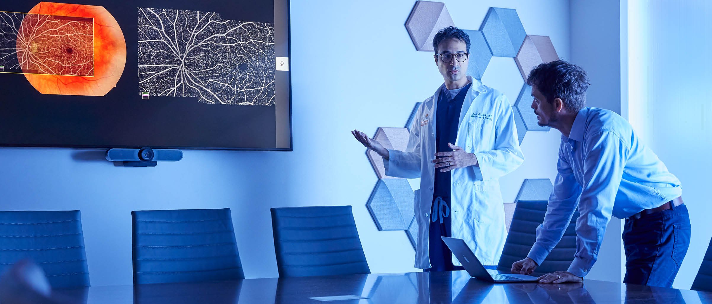
Retinal cameras—small, low-power microscopes commonly found in eye doctor offices—use high-resolution imaging to take pictures of the inside of the eye. “Because the retina is directly connected to the brain, we think we can use this as a proxy to see what’s happening in the brain,” explains Giancardo. The team’s goal is to develop an algorithm that examines three different types of images to evaluate what is happening to the vasculature of the retina.
“This could have a significant impact on morbidity and mortality,” he says. “And we have the expertise in stroke, ophthalmology, and machine learning to do this.”
The project is a collaboration between Giancardo; Sunil A. Sheth, MD, vascular and interventional neurologist at UTHealth Neurosciences and McGovern Medical School; Charles Green, PhD, Associate Professor in the Department of Pediatrics at McGovern Medical School; and Roomasa Chana, MD, an ophthalmologist at Baylor College of Medicine. The team garners additional support from UTHealth Institute for Stroke and Cerebrovascular Disease, which fosters collaborations in stroke research among the schools of UTHealth. This includes Sean I. Savitz, MD, and Amanda Jagolino-Cole, MD, who provide care through UTHealth Neurosciences.
While the research grant specifically focuses on strokes in space—where zero gravity and prolonged space radiation increase the risk of cardiovascular disease and stroke in even the healthiest individuals—it has implications for rural areas of the United States and any place that lacks immediate access to diagnostic brain imaging equipment.
But the team’s research doesn’t stop there. Giancardo and Sheth recently helped create an algorithm that identifies large vessel occlusions, a specific type of ischemic stroke that blocks blood flow of one of the main arteries in the brain and may account for up to one-third of ischemic strokes. Using a dye injected into the body and CT imaging—instead of CT perfusion imaging, which evaluates blood flow—the algorithm can determine within a minute if a patient is suffering from a large vessel occlusion and notify the patient’s health care team. The algorithm even helps determine if the patient is eligible for endovascular thrombectomy, a minimally invasive treatment where a tiny catheter is threaded through the blood vessel to remove the clot.
“When physicians see that someone has a large vessel occlusion, they send them to a large hospital for CT perfusion imaging to determine this,” explains Giancardo. When this happens, precious time is lost. “We wanted to see if we could create a proxy for CT perfusion using just CT imaging, which is readily available at smaller hospitals.
Giancardo’s team at the Center for Precision Health at the School of Biomedical Informatics has established a pipeline to translate research at UTHealth into something that can benefit the patients immediately. Philanthropy can accelerate discoveries like this by providing seed funding to initiate high-risk, high-reward projects and garner the data necessary to apply for larger funding sources.
“For most federal funding agencies, the turnaround time is six months if you are lucky,” says Giancardo. “That is a very long time, especially in my world where developments happen super quickly.”
And like the experts say: Time is brain.
Busting the clot
During a thrombectomy, a tiny catheter is threaded through the arteries to physically remove the clot and restore blood flow. Although lifesaving in many cases, the standard treatment for an ischemic stroke caused by a large vessel occlusion may provide no benefit for more than a third of patients - a statistic that does not sit well with P. Roc Chen, MD, a physician-scientist at UTHealth Neurosciences.
“We have a significant opportunity to improve acute stroke treatment,” Chen says.
A substantial debate among stroke treatment physicians involves whether patients have better outcomes under general anesthesia or conscious sedation. While some retrospective studies favor conscious sedation, they suffer from a research shortfall known as selection bias: Because doctors tend to place the most gravely ill stroke patients—who are less likely to have positive outcomes— under general anesthesia, this method may falsely appear to lead to poorer results.
Working with the UTHealth Institutional Review Board to swiftly enroll patients when they arrive at hospitals, Chen launched a multi-center randomized study funded by philanthropy and industry grants to seek a clearer answer for the future standard of care.
“This could be a milestone for other stroke studies,” he says. “I hope it will help resolve the debate.”
Chen has also set his sights on cerebral vasospasm induced by ruptured brain aneurysms, weak spots in brain blood vessels that balloon and burst. Nearly half of patients with ruptures will die, but many not from the rupture itself; when the leaked blood breaks down, it irritates nearby blood vessels, causing cerebral vasospasm leading to stroke. By analyzing data from past rupture treatments, Chen found that a combination of three medications helped improve patient outcomes, so he launched a multicenter clinical trial to evaluate this method’s effectiveness.
Even after successful treatment, however, patients who suffer an aneurysm must follow up with physicians for periodical angiograms—an invasive procedure that many patients skip because of the high costs—to check for reoccurrence. Chen is working on a project funded by the National Institutes of Health to determine if computer algorithms can analyze simple x-ray images to determine the risk of an aneurysm striking again. “
If patients only need to have an x-ray, this could significantly improve follow-up rates,” he says. “The easier we can make the process, the more lives we can save.”