
Resting state functional connectivity patterns as biomarkers for treatment in patients with depression


Brain images revealed widespread dysfunction of resting-state effective connectivity in people with depression, according to research published in the Journal of Affective Disorders by researchers at UTHealth Houston.
Researchers hope that the research can led to earlier diagnosis, as well as predicting which patients might respond better to a particular medication.
“We don’t have any objective way to say that someone is depressed based on a biological marker,” said Sudhakar Selvaraj, MD, PhD, associate professor and director of the Depression Research Program at the Louis A. Faillace, MD, Department of Psychiatry and Behavioral Sciences with McGovern Medical School at UTHealth Houston. “We make the diagnosis, and pick a treatment based on the patient’s interview and our clinical assessment. Currently, we don’t have any markers to say to our patients, this is happening in your brain, so this is why you’re getting this treatment.”
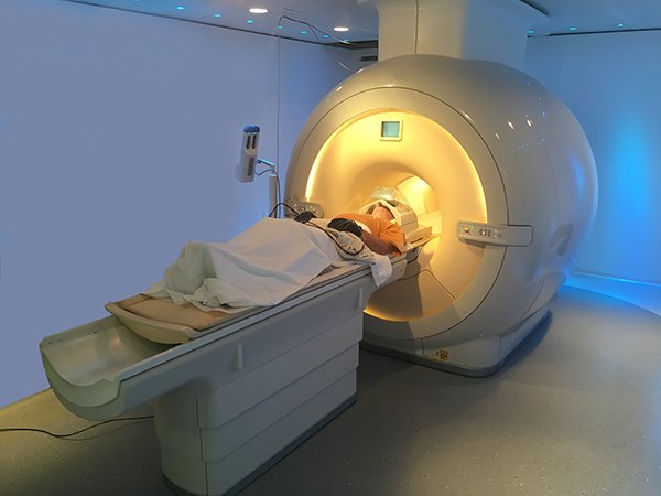
Researchers led by Selvaraj and first author, Dana DeMaster, PhD, assistant professor of pediatrics with McGovern Medical School and Children’s Learning Institute at UTHealth Houston, used a functional MRI (fMRI) to analyze and measure brain activity during the resting state in 34 drug-free patients with major depressive disorder. This research targeted functioning of the default mode network in the brain because it is engaged during periods of rest and plays a role in self-referential processing. Resting-state fMRI results showed the default mode network was strongly connected in people with depression. Connections were also evident from the default mode network to the central executive network which supports cognitive functioning such as decision-making. Importantly, for the salience network, which allows environmental stimuli to trigger the brain to cycle out of internal mental processes, connections to either the default mode network or central executive network were highly reduced in this depressed sample.
“When we ran that analysis, what we found is that connections between networks were not as strong as you would expect,” DeMaster said. “We inferred from that information that balancing physiological reaction to all of the stimuli in our environment might not be as strong as in patients with major depression. And that is consistent with previous work, but we were able to show that in terms of regions signaling to one another, not just the synchrony with one another.”
After the baseline fMRI, patients were treated for six weeks with escitalopram, a standard first-line antidepressant. After the six weeks of treatment, the data from the fMRI revealed that 21 (62%) patients were classified as responders to escitalopram.
“Using a brain scan allowed us to understand what is happening to the brain when someone is feeling depressed and how medication changes the brain connections to improve your mood and symptoms of depression,” Selvaraj said. “We hope we can objectively show the patient what is happening in their brain and how the treatment is changing the brain activity, and that would be a big relief for the patient.”
This study was a small open-label study, meaning the patient and doctor knew what antidepressants they were getting. The next step in this research will be to look into whether the same model can be applied and predict the same kind of pattern in a larger sample with a placebo.
“Brain activity can change if you know what treatment you’re getting because your expectation can change the brain scans, so we wanted to address that part in the bigger analysis,” Selvaraj said. “We will look into studies where there is a placebo compared with the antidepressant. So, the bottom line is we want to replicate these results in a larger sample, and then see whether this model can be useful in predicting people responding to the antidepressant.”
Beata R. Godlewska, MD, with the Department of Psychiatry, Medical Sciences Division, University of Oxford, and Oxford Health NHS Foundation Trust was a co-author on the study.
Media inquiries: 713-500-3030