
Translational Technologies and Resources (Core Labs)
Core Laboratories
The CCTS works with many different laboratories to conduct cutting-edge medical research. They connect researchers with these labs to foster collaboration and clinical translation. The core labs are listed below.
Atomic Force Microscopy Lab
Location: 3SCRB6.3728
Director: Ana Maria Zaske, PhD - [email protected] - 713-486-5418
Atomic force microscopy (AFM) is a technique for characterizing surfaces and can reproduce topological images under physiological-like conditions. AFM is a noninvasive technique base on bio-sensing. Scanning can be performed in air or in a liquid environment, and a range of samples, from living cells down to single molecules, can be imaged. AFM is also an attractive tool for studying the endocytosis of nano-vectors and the systemic response to biological processes. An area of major interest is determining the stiffness of a sample (elastic modulus). The elasticity of the cell membrane can vary between cell types as a function of growth, differentiation, disease, or treatment. Non-cellular structures can also be imaged. The core uses a Bioscope II Scanning Probe Microscope specifically designed to address the needs of biological and medical investigations. It has unprecedented compatibility with optical microscopy. All three axes of the instrument are equipped with high-resolution capacitive sensors; this allows imaging at the nanoscale level and in liquids. The microscope requires minimal sample preparation.
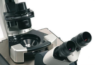
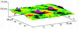
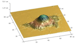
Left: BioScope II scanner (Bruker Inc); Middle: 3D AFM image of liposomes during internalization; Right: 3D AFM image of a HeLa cell
Pre-Clinical CT Core Facility
Location: MSB 5.302
Director: Delia Danila, PhD - [email protected] - 713- 486-6531
This core laboratory has a specialized CT system for rapid high-resolution imaging of objects up to the size of large rabbits. The facility utilizes a GE eXplore Ultra Pre-Clinical CT Scanner (GE Healthcare, London, ON) for high-resolution (150-micrometer isotropic voxel size) cone-beam CT engineered for small-animal imaging (maximum 14-cm diameter field of view).
The system permits up to 1-second gantry rotation times, and X-rays are produced by a variable voltage X-ray tube capable of energies from 70 to 140 kVp. This provides high-quality images with contrast-to-noise, resolution, and dose performance optimized for pre-clinical imaging. This imaging modality can facilitate studies in many disciplines, including orthopedics, pulmonary, cardiovascular, gastrointestinal, genitourinary, oncology, and others. The specific 2D and 3D analyses provided by this technology include vascular analysis, bone analysis, bone mineral density (BMD), volumetric measurements, and contrast agent development. Please contact Dr. Danila for more information or to submit an imaging request.
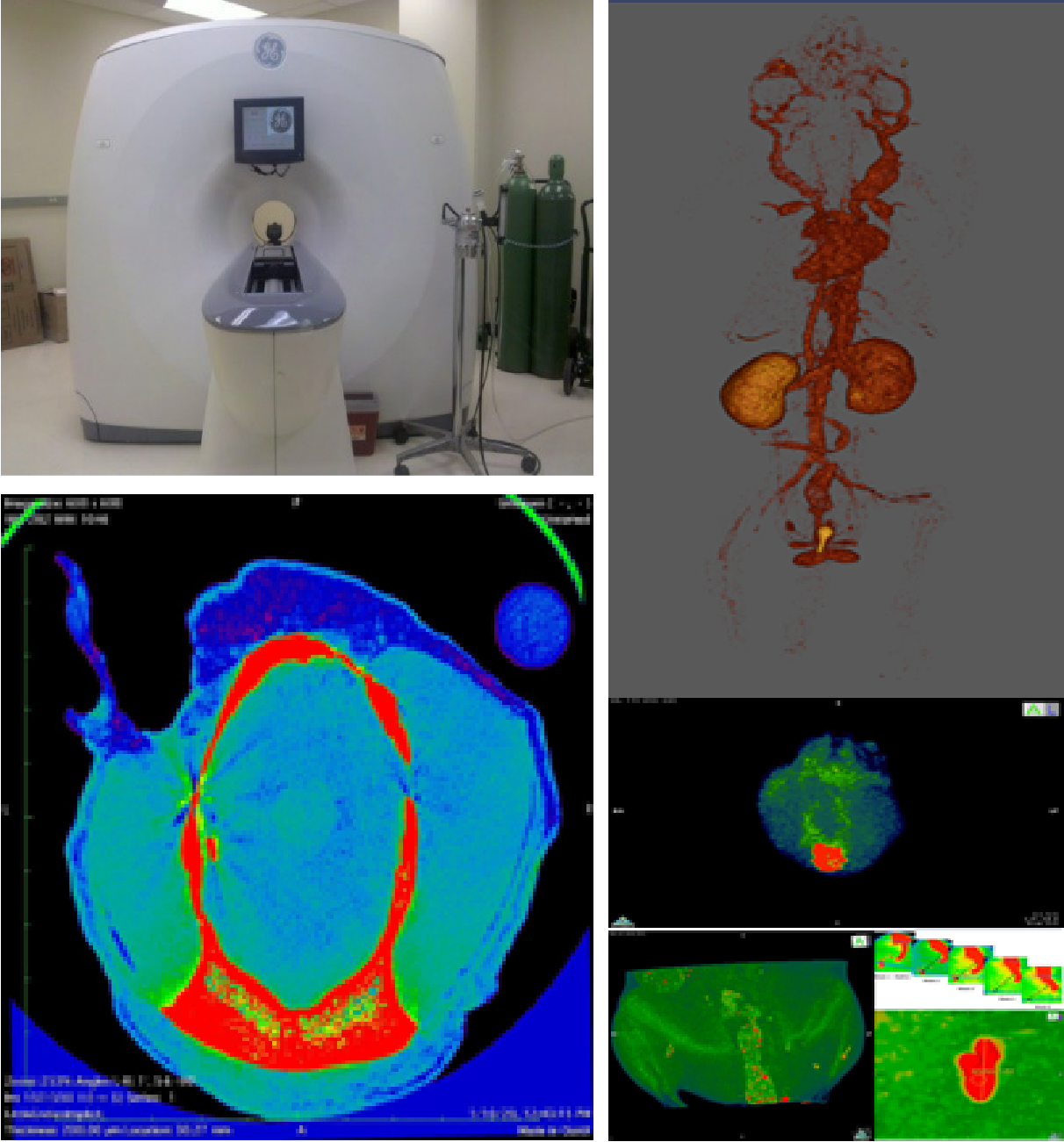
Top left: 4-GE eXplore Ultra; Bottom left: Scan of brain taken with the GE eXplore Ultra; Left: images taken at the CT lab.