In the future, portable retina imaging systems could be the key to diagnosing acute strokes
Collaboration between multiple UTHealth Houston schools and departments produced evidence of the feasibility of such a system by training machine learning models

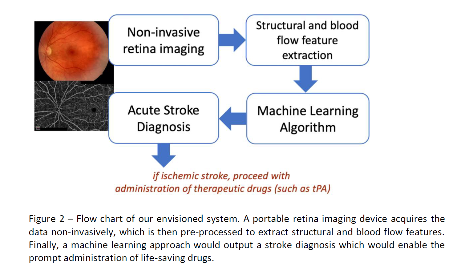
Stroke is one of the leading causes of death and disability worldwide. The time it takes from symptom onset, travel to an emergency center, diagnosis with imaging, to treatment can waste precious minutes. The longer it takes for a patient to receive treatment, the risk of cognitive and physical decline following the stroke increases. The time could be reduced with a prompt diagnosis during patient transportation to the hospital. A portable retina imaging system could enable this by measuring vascular information and blood perfusion in the retina and, due to the homology between retinal and cerebral vessels, infer if a cerebral stroke is underway.
The team at the School of Biomedical Informatics (Luca Giancardo, Ivan Coronado, Juntao Yan), McGovern Medical School (Amanda Jagolino-Cole, Sunil Sheth, Rania Abdelkhaleq, Charles Green, Sergio Salazar Marioni), Institute for Stroke and Cerebrovascular Diseases, and in collaboration with the University of Wisconsin (Roomasa Channa), implemented a project, made possible due to a grant from the Translational Research Institute for Space Medicine (NNX16AO69A), to shorten the time to treatment using alternative tools.
Why Is Space Medicine Involved?
The interest from space medicine comes from the documented increased risk of cerebrovascular disease associated with prolonged exposure to radiation in space. This can greatly increase the risk of stroke during deep space missions, and in space there is no room for CT or MRI brain imaging onboard spacecraft. So how would they detect a stroke in space? The answer may be retinal imaging.
How Does It Work?
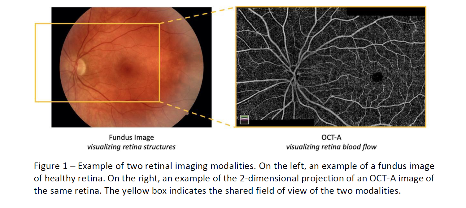
In their preliminary results, researchers were able to train a simple machine learning approach to distinguish controls from acute stroke subjects within four days from a stroke event using OCT-A images. Optical coherence tomography (OCT) is a noninvasive imaging method that uses reflected light to create pictures of the back of your eye. Measurements captured from these images can capture the distribution of blood flow at different layers. Their vision is to create a portable system that can diagnose stroke everywhere from ambulances to deep space missions.
Read More here and see the dashboard for the algorithm here