
Luca Giancardo, PhD
Associate Professor at the Center for Precision Health, School of Biomedical Informatics (SBMI), University of Texas Health Science Center at Houston (UTHealth)
Luca Giancardo is an Associate Professor at the Center for Precision Health, School of Biomedical Informatics (SBMI), University of Texas Health Science Center at Houston (UTHealth) with co-appointments at the McGovern Medical School and the Institute for Stroke and Cerebrovascular Diseases, UTHealth.
He is a computer scientist with extensive experience in image analysis and machine learning. I have worked on developing new machine learning-based methodologies to discover computational biomarkers from patterns in biomedical data such as optical images, magnetic resonance imaging, X-rays, computer tomography, laboratory animal videos or tracking devices. His work has been applied to a number of biomedical applications, such as stroke diagnosis, diabetic retinopathy screening or neurodegenerative disease tracking and successfully translated to industry with two startups based on his methods. One, Hubble Telemedical, was acquired by Welch Allyn in 2015, another, nQ-Medical has raised multimillion dollars in investment. He has authored/co-authored more than 60 peer-reviewed articles (H-index 20) which were featured by news outlet such as MIT Technology Review, Smithsonian magazine and others. He has received multiple awards, including the prestigious 100k Singapore Challenge (judging panel composed by Nobel Prize and Millennium Technology Prize winners).
He was awarded competitive grants from NIH (R01), Translational Research Institute for Space Health, Michael J Fox Foundation and private companies.
Research News
Quantification of infarct core signal using CT imaging in acute ischemic stroke
In stroke care, the extent of irreversible brain injury, termed infarct core, plays a key role in determining eligibility for acute treatments, such as intravenous thrombolysis and endovascular reperfusion therapies. Many of the pivotal randomized clinical trials testing those therapies used MRI Diffusion-Weighted Imaging (DWI) or CT Perfusion (CTP) to define infarct core. Unfortunately, these modalities are not available 24/7 outside of large stroke centers. As such, there is a need for accurate infarct core determination using faster and more widely available imaging modalities including Non-Contrast CT (NCCT) and CT Angiography (CTA).
New NASA Grant to Detect Strokes in Space
An untreated stroke event would be destructive for a human deep space exploration mission. Increased cerebrovascular disease risk has been documented after prolonged exposures to ionizing radiations on Earth. Astronauts on deep space exploration missions will be exposed to galactic cosmic rays and solar particles for 30 months, which can lead to accelerated vascular injury likely increasing their risk of stroke.
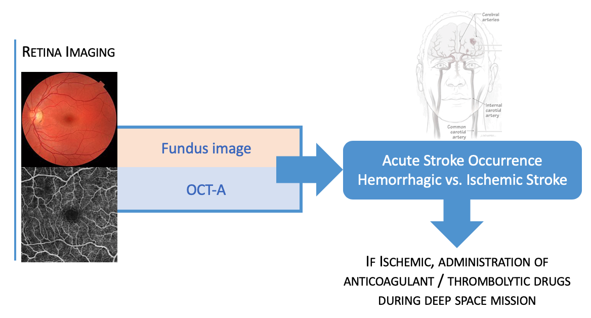
On Earth, ischemic strokes can be successfully treated with anticoagulant or thrombolytic drugs if the event is rapidly diagnosed and the type of stroke (ischemic versus hemorrhagic) is rapidly identified with Computer Tomography (CT) or Magnetic Resonance Imaging (MRI) brain imaging. However, these brain imaging capabilities do not exist in space and alternative robust means of diagnosing and classifying stroke are needed. Due to the homology between retinal and cerebral vessels, and the ease with which retinal images can be acquired non-invasively, retinal images have been studied as a markers for cerebrovascular events. Retinal vascular abnormalities associated with incident stroke include arteriolar narrowing and reduced vessel fractal dimension. We propose to use a combination of color fundus photos and optical coherence tomography angiography (OCT-A) images to identify stroke events and stroke type, effectively acting as a proxy for brain imaging. These imaging modalities are non-invasive and deployable in currently existing technologies for deep space missions. We will adapt our automated interpretable image-based deep learning algorithm to identify stroke and stroke type from retinal vascular images, enabling an automated life-saving tool usable on a deep space exploration mission.
In addition to Dr. Giancardo the research team is composed of: Roomasa Channa, MD (co-I). Baylor College of Medicine, Retina Specialist with experience in identifying retinal abnormalities and their connections to overall health assessment. Sunil A. Sheth, MD (co-I), UTHealth McGovern Medical School. Stroke neuroimaging specialist with experience in stroke dataset creation for machine learning development. James Grotta, MD a vascular neurologist at the UTHealth McGovern Medical School and Sean Savitz, MD, the director of UTHealth Institute for Stroke and Cerebrovascular Disease will have an advisory role for data acquisition and interpretation.
Education & Training
Doctorate
Oak Ridge National Laboratory, US and University of Burgundy, France
Postdoc
The Italian Institute of Technology, Italy
Research Fellowship
The Massachusetts Institute of Technology, Boston, MA